Questions? 800-523-5874 | [email protected]
- Prepmaster™ Specimen Preparation Robot
- TEM Grids
- TEM Window Grids
- Omniprobe Nanomanipulation Systems
- K-kit Wet "Liquid" TEM Kit
- Specimen Mounts
- SEM Specimen Holders
- Index and Finder SEM Grids
- SEM for Forensics
- SEM Sample Preparation Station Materials
- Cryogenic Personal Protection Equipment
- Cryo Dewars & Flasks
- Cryogenic Grids & Accessories
- Cryogenic Vials & Racks
- Cooling Chambers & Ice Baths
- Prepmaster™ Specimen Preparation Robot
- Laboratory Microwave Ovens
- LYNX II Automated Tissue Processor
- EMS Poly III
- Microtomes
- Tissue Slicers
- Rapid Immersion Freezer
- Heaters & Chillers
- SEM Cooling Stage
- Glow Discharge Systems
- Sputter Coaters & Carbon Coaters
- Stages
- Freeze Dryers
- Critical Point Dryers
- Cryo-SEM Preparation System
- Specimen Transfer Systems
- Decontaminators
- Desiccators
- Centrifuges
- Dry Baths
- Stirrers, Hot Plates
- Vortexers & Magnetic Mixers
- Rotators & Rockers
- Ovens & Incubators
- Vibration Isolation
- Air Sampling
- Vacuum Pumps
Product Specials

Dear Fellow Researcher,
We are pleased to offer all of our loyal customers these special programs and discounts to help in your scientific research needs.
 Special Offer...
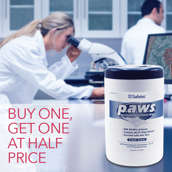
Buy one P.A.W.S. Canister Dispenser, get one 1/2 off
Electron Microscopy Sciences is pleased to offer a special discount for P.A.W.S. Personal Antimicrobial Wipes available while supplies last.
Discount applies to Canister Dispenser version only, catalog number 70576-50. Discount will only be shown in cart when you add 2 of the applicable product. Limit of 1 P.A.W.S. special discount per order.
Precision Diamond Wafering Blades - While supplies last... receive 20% off select blades! Call to order.
These bonded blades are constructed of an inner metal core and an outer rim; this rim is bonded with diamond particles. These blades come in high and low concentrations of diamond particles to handle various sectioning requirements. 20% discount applies to EMS Catalog numbers:
| 50265-FH3 | 50265-FH4 | 50265-MH4 | 50265-CL4 | 50266-FH4 |
| 50266-MH4 | 50266-CH4 | 50267-MH4 | 50265-MH5 | 50265-CH5 |
| 50265-CH6 | 50265-CL6 | 50265-MH7 | 50265-04 | 50266-04 |
| 50266-05 | 50266-06 | 50266-07 | 50267-06 |
EMS Grids - For a limited time, buy 10 EMS Grid vials, get 3 EMS Grid vials of equal or lesser value free!
A reliable support specimen grid source. Features: well-defined grid bars, maximum open area, and a matte/shiny side. Each grid is individually inspected. Newly introduced are copper grids with palladium plating. This plating offers better grid strength and avoids tarnishing. Also new are Copper-Rhodium (Cu-Rh) grids in mesh sizes 200 and 300.
Mic-Fi / Visio-tek Digital Wi-Fi/USB Microscopes - Order a new Mic-Fi / Visio-tek Digital Wi-Fi/USB Microscopes and receive 10% off.
Mic-Fi / Visio-tek is a line of versatile, portable digital microscopes with Wi-Fi transmission. Every device can be connected to iOS and Android smartphones or tablets through a specific Mic-Fi / Visio-tek App or through a Wi-Fi/USB connection to computers and laptops (Windows or Mac). They break the conception of traditional microscopes to realize the following functions: measurement, conservation, copy and transfer of images and video, which are difficult for a traditional microscope. They are small, easy to operate, light, and portable. They can be used with all the most popular smartphones, tablets and even with PCs.
PP3010 Cryo-SEM Preparation System - Order a PP3010 and receive a $2500 credit towards the purchase of any of our EMS line of products.
The PP3010 is a highly automated, easy to use, column-mounted, gas-cooled cryo preparation system suitable for most makes and models of SEM, FE-SEM and FIB/SEM. The PP3010 has all the capabilities to rapidly freeze, process and transfer specimens. The cryo preparation chamber is turbomolecular pumped and includes tools for cold fracturing, controlled sublimation and specimen coating. The specimen can then be transferred onto a highly stable SEM cold stage for observation. Cold trapping in the cryo preparation chamber and SEM chamber ensures the whole process is frost free. Specimen process times are typically between five and ten minutes.
Aurion UltraSmall ImmunoGold Reagents and Conventional ImmunoGold Reagents - Order any of our Ultrasmall Reagents and receive a $25 gift certificate good towards the purchase of any of our EMS line of products.
Reduction of the gold particle size provides Ultra Small Immunogold Reagents with fundamentally different characteristics when compared with conjugates built around larger particles. While the Conventional Immunogold Reagents can be thought of as particles coated with proteins, Ultra Small Immunogold Reagents are proteins coated with one or more gold particles. With this structure, both the overall size of the conjugates, as well as steric hindrance are decreased.
Innovative and Versatile Sputter Coaters and Carbon Evaporator- For all Electron Microscopy Applications: SEM, high resolution FESEM and TEM - Buy a new unit today and get a $500 credit towards the purchase of any accessories we offer.
The Q150R Plus, the Q150T Plus and the Q300 Plus Series offer a complete range of stand alone carbon coaters, sputter coaters, Sputter/Carbon coaters in one. We offer low-cost, rotary-pumped systems for depositing non-oxidizing metals - such as gold (Au) and platinum (Pt) - and also turbomolecular-pumped models, suitable for oxidizing and non-oxidizing metals - such as chromium (Cr). Large chamber models are available for specimens up to 8"/200mm diameter, and all sputter coaters can be fitted with carbon evaporation attachments.
- Q150R Plus - EMS Series of Rotary Pumped Modular Coating Systems
- Q150T Plus - Turbo-Pumped Sputter Coater/Carbon Coater
- Q150V Plus - EMS Series of Ultra-Fine Coating Systems
- Q300T D Plus - Dual Target, Large Chamber, Turbo-Pumped Sputter Coater
- Q300T T Plus - Triple Target, Large Chamber, Turbo-Pumped Sputter Coater
NIGHTSEA® Stereo Microscope Fluorescence Adapter - Adapt your existing lab stereo microscopes for fluorescence - Buy a NIGHTSEA SFA and receive a $50 gift certificate for any of our consumables and supplies.
The NIGHTSEA Stereo Microscope Fluorescence Adapter adapts just about any stereo microscope (dissecting microscope) for fluorescence with no modification to the microscope itself. The modular design lets you easily switch between several different excitation/emission combinations to work with a variety of fluorescent proteins and other fluorophores. There are now six different excitation/emission combinations available, plus white light.
DiATOME Diamond Knives - For US Customers Only
- The Ultra Exchange Program
- Exchange any old diamond knife from any manufacturer for a brand new Ultra diamond knife and pay almost half the price of a new knife. No matter what size you trade in, you may choose any size DiATOME Ultra knife at the reduced price.
- The Multiple Purchase and Giveaway Program
- For your convenience we have set this program up in three different ways:
Buy 2 Ultra room temperature or cryo knives at full price, get a third knife 50% off OR
Buy 1 Diamond knife at full price, get a second knife at the Exchange price OR
Resharpen 2 knives, get a third knife resharpened for free. - The Histo Knife Regular and Jumbo Exchange Program
- Exchange any old diamond knife from any manufacturer for a brand new Histo diamond knife (Regular or Jumbo) and pay a discounted price for a new knife. No matter what size you trade in, you may choose any size Diatome Histo knife.
- The DiATOME Rebate Program
- This program allows you to purchase any new DiATOME Diamond Knife at its regular price and receive a $250.00 coupon towards the purchase of chemicals, accessories and supplies from Electron Microscopy Sciences.