Questions? 800-523-5874 | [email protected]
- TEM Grids
- Prepmaster™ Specimen Preparation Robot
- TEM Window Grids
- Omniprobe Nanomanipulation Systems
- K-kit Wet "Liquid" TEM Kit
- Specimen Mounts
- SEM Specimen Holders
- Index and Finder SEM Grids
- SEM for Forensics
- SEM Sample Preparation Station Materials
- Cryogenic Personal Protection Equipment
- Cryo Dewars & Flasks
- Cryo-EM Grids & Grid Storage
- Cryogenic Vials & Racks
- Cryo-EM Vitrification Supplies
- Prepmaster™ Specimen Preparation Robot
- Laboratory Microwave Ovens
- LYNX II Automated Tissue Processor
- EMS Poly III
- Microtomes
- Tissue Slicers
- Heaters & Chillers
- SEM Cooling Stage
- Glow Discharge Systems
- Sputter Coaters & Carbon Coaters
- Stages
- Freeze Dryers
- Critical Point Dryers
- Cryo-SEM Preparation System
- Specimen Transfer Systems
- Decontaminators
- Desiccators
- Centrifuges
- Dry Baths
- Stirrers, Hot Plates
- Vortexers & Magnetic Mixers
- Rotators & Rockers
- Ovens & Incubators
- Vibration Isolation
- Air Sampling
- Vacuum Pumps
EMS Technical Data Sheets

UranyLess – Protocols of Use: Negative Staining
EMS Catalog #22409
Negative staining is a very useful technique in electron microscopy. It allows characterization of isolated particles of morphology as bacteria, virus, protein, nanoparticles, liposomes, exosomes, etc.
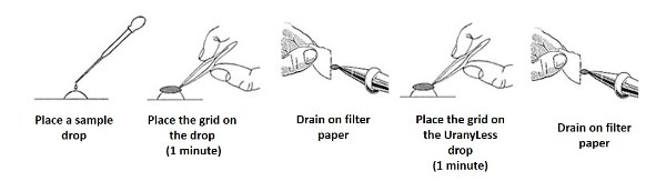
Staining Protocol #1:
- On a piece of parafilm or any other hydrophobic carrier, place a drop of your solution (~ 10µl) and a UranyLess drop.
- Using our fine tweezers, place your sample drop on a formvar-carbon coated grid. for about 1 minute.
- Blot your grid using filter paper.
- Place your grid on the UranyLess solution for 1 minute.
- Blot, let it dry for 5 minutes and observe under the microscope.
Technical Tip:
- If the staining is too intense, wash with room temperature water for 1 minute.
Staining Protocol #2 (Debra M. Townley, Baylor College of Medicine):
- One drop of suspension of 300 mesh formvar coated grids – 30 minutes
- Wick away excess suspension with paper arrow (Watman No.1)
- Place one drop of Uranyless on the grid before it is completely dry
- Stain for 3 minutes
- Wick away excess stain with paper arrow (Watman No.1)
- Allow the grids(s) to dry completely before examination on the TEM
- Times can be altered (suspension time, stain time) to suit the "look" you desire