Questions? 800-523-5874 | [email protected]
- TEM Grids
- Prepmaster™ Specimen Preparation Robot
- TEM Window Grids
- Omniprobe Nanomanipulation Systems
- K-kit Wet "Liquid" TEM Kit
- Specimen Mounts
- SEM Specimen Holders
- Index and Finder SEM Grids
- SEM for Forensics
- SEM Sample Preparation Station Materials
- Cryogenic Personal Protection Equipment
- Cryo Dewars & Flasks
- Cryo-EM Grids & Grid Storage
- Cryogenic Vials & Racks
- Cryo-EM Vitrification Supplies
- Prepmaster™ Specimen Preparation Robot
- Laboratory Microwave Ovens
- LYNX II Automated Tissue Processor
- EMS Poly III
- Microtomes
- Tissue Slicers
- Heaters & Chillers
- SEM Cooling Stage
- Glow Discharge Systems
- Sputter Coaters & Carbon Coaters
- Stages
- Freeze Dryers
- Critical Point Dryers
- Cryo-SEM Preparation System
- Specimen Transfer Systems
- Decontaminators
- Desiccators
- Centrifuges
- Dry Baths
- Stirrers, Hot Plates
- Vortexers & Magnetic Mixers
- Rotators & Rockers
- Ovens & Incubators
- Vibration Isolation
- Air Sampling
- Vacuum Pumps
EMS Technical Data Sheets

Pyrolytic Graphite Stripper Film
EMS Catalog #76040-05, 76040-10, 76041-05, 76041-10
Introduction
Over years of extensive research, high-energy radionuclide production with the incorporation of cyclotrons has gradually become very well known for researchers, scientists, microscopists, and other professionals alike. An abundance of new applications for PET imaging has quickly surfaced, moving from nuclear cameras with coincidence detection to mobile and fixed PET and PET/CT scanners. Did you know that PET imaging is conducted in well over 2,000 different sites, using said scanners? Remarkable!
With the unfortunate, rapid increase in various cases of cancer and diseases, the vital need for precise diagnosis is more evident than ever.
- Highest thermal stability
- Highest purity
- Highest uniformity
Carbon foils
Carbon foils are thin films used to strip electrons to produce a short-lived radionuclide that, when injected into a patient, enables the PET imaging process. It is the carbon foils that make high energy research and radionuclide production possible.
Please note that the lifespan of the stripper foil depends on the type of carbon used and the means of application.
In an examination in which free-standing Pyroid pyrolytic graphite stripper foils were compared, alternative arc-evaporated carbon and PCG graphite foils were used in an ISO Certified Laboratory Testing Facility.
Results of the examination: Field Emission Scanning Electron Microscopy (FESEM)
FESEM was utilized to examine the free-standing stripper foil samples from general applications for Pyroid PG, and commercially available arc-evaporated, PCG foils.
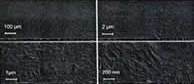
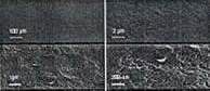
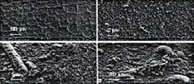
Please refer the following figures below, which compare FSEM to various Pyroid PG stripper foils:| Figures 1 and 2 compare FSEM results of Pyroid PG stripper foil with an areal density of 400µg/cm2 with an arc-evaporated stripper foil with an areal density of 250µg/cm2. | ||
 |
 |
|
| Figure 1: Pyroid Pyrolytic Foil Areal Density 400µg/cm2 | Figure 2: Arc-Evaporated Foil Areal Density 250µg/cm2 | |
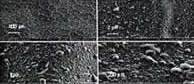
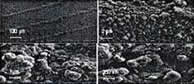
| Figures 3, 4 and 5 compare FSEM results of Pyroid PG stripper foil with an area density of 1,000µg/cm2 with an arc-evaporated stripper foil and PCG foil with an areal density of 1,000 µg/cm2. | ||
 |
 |
 |
| Figure 3: Pyroid Pyrolytic Foil Areal Density 1,000µg/cm2 | Figure 4: Arc-Evaporated Foil Areal Density 1,000µg/cm2 | Figure 5: PCG Foil Areal Density 1,000µg/cm2 |
Note: In each of the figures above, the Pyroid PG foils yielded the most consistent physical appearance. When compared to commercial samples, features as such are generally unseen. Also noted, on the contrary, in non-uniform appearances, foil expansion can occur. Foils are generally mounted with the purpose of localizing the central part. Any surfaces that appear raised in the arc-evaporated and PCG foils under FSEM are usually hollow and indicate stress under irradiation.
X-Ray Diffraction (XRD)/Thermo gravimetric Analysis/Differentia Thermal Analysis (TGA-DTA)
When under observation, Pyroid PG foils tend to undergo typical conical crystalline pyrolytic graphite structure with patterns in its uniformity, consistency, and lack of non-graphite species. This, then, contrasts with the arc-evaporated foils that show evidence of carbon-like formation on the foil.
Furthermore, the PCG foils contain about 25% non-graphitic, low temperature organic contamination.
Note: A foil will quickly be destroyed if its temperature rises to the point at which evaporation becomes of significance or if melting begins.