Questions? 800-523-5874 | [email protected]
- TEM Grids
- Prepmaster™ Specimen Preparation Robot
- TEM Window Grids
- Omniprobe Nanomanipulation Systems
- K-kit Wet "Liquid" TEM Kit
- Specimen Mounts
- SEM Specimen Holders
- Index and Finder SEM Grids
- SEM for Forensics
- SEM Sample Preparation Station Materials
- Cryogenic Personal Protection Equipment
- Cryo Dewars & Flasks
- Cryo-EM Grids & Grid Storage
- Cryogenic Vials & Racks
- Cryo-EM Vitrification Supplies
- Prepmaster™ Specimen Preparation Robot
- Laboratory Microwave Ovens
- LYNX II Automated Tissue Processor
- EMS Poly III
- Microtomes
- Tissue Slicers
- Heaters & Chillers
- SEM Cooling Stage
- Glow Discharge Systems
- Sputter Coaters & Carbon Coaters
- Stages
- Freeze Dryers
- Critical Point Dryers
- Cryo-SEM Preparation System
- Specimen Transfer Systems
- Decontaminators
- Desiccators
- Centrifuges
- Dry Baths
- Stirrers, Hot Plates
- Vortexers & Magnetic Mixers
- Rotators & Rockers
- Ovens & Incubators
- Vibration Isolation
- Air Sampling
- Vacuum Pumps
EMS Technical Data Sheets

Stereo Microscope Fluorescence Aided Dissection of GFP Labeled Structures
EMS Catalog #SFA
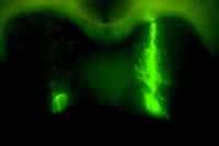
Region of mouse brain into which EGFP-tagged lentovirus vector has been injected. © Marina Wolf, Rosalind Franklin University.
Green Flourescence Protein (GFP)-labeled dorsal striatum in mouse brain. © NIGHTSEA/Charles Mazel. Sample photographed at laboratory of Stefano Vicini, Georgetown University.
The most common application of NIGHTSEA lights for researchers using fluorescent proteins is in sorting out which members of the next generation are fluorescent and which are not. Whether working with mouse pups, Drosophila larvae, zebrafish, or other organisms, the lights make it easy to see which offspring have inherited the fluorescence trait and which have not.
Some researchers are going beyond just identifying labeled subjects and using fluorescence to actively aid in extracting Green Flourescence Protein (GFP)-tagged structures. In one case the researchers needed to extract only the GFP-tagged dorsal striatum from within mouse brains. They likened this to "isolating a lump of oatmeal from within a larger lump of oatmeal". When they switched from doing the dissection in white light to using the NIGHTSEA flashlight and glasses they could easily see which portion of the brain to target. It made the dissection both faster and more accurate.
In another case the researchers needed to punch tissue from the nucleus accumbens of a mouse for subsequent biochemical analysis. Being able to see the fluorescence in real time made it easy to target the right structure. (See - Xuan Li and Marina E. Wolf, 2011. Visualization of virus infected brain regions using a GFP-illuminating flashlight enables accurate and rapid dissection for biochemical analysis. J. of Neuroscience Methods, Vol. 201, Issue 1, pp. 177-179.)
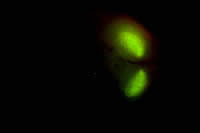
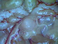
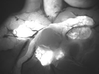
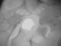
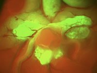
Dr. Xin Lu, a researcher at the MD Anderson Cancer Center in Houston, contributed some nice images of his work with Green Flourescence Protein (GFP)-tagged tumors in a universally red-fluorescent (RFP) mouse. Dr. Lu purchased the NIGHTSEA Stereo Microscope Fluorescence Adapter to add to his Nikon SMZ745 stereo microscope so that the tumor would really stand out, making it easier to dissect. The photos below show the image (1) in white light, (2) using the Royal Blue excitation/emission set to capture the green fluorescence, (3) using the Green excitation/emission set to capture the red fluorescence, and finally (4) a color composite of the green and red channel images.
 |
 |
|
| (1) White light – Green Flourescence Protein (GFP) tumor in RFP mouse. Exposure made with white light. © Xin Lu, MD Anderson Cancer Center | (2) Green fluorescence – GFP tumor in RFP mouse. Exposure made to highlight the green fluorescence. © Xin Lu, MD Anderson Cancer Center |
 |
 |
|
| (3) Red fluorescence – GFP tumor in RFP mouse. Exposure made to highlight the red fluorescence. © Xin Lu, MD Anderson Cancer Center |
(4) Fluorescence color composite – GFP tumor in RFP mouse. Composite of green and red fluorescence. © Xin Lu, MD Anderson Cancer Center |