Questions? 800-523-5874 | [email protected]
- TEM Grids
- Prepmaster™ Specimen Preparation Robot
- TEM Window Grids
- Omniprobe Nanomanipulation Systems
- K-kit Wet "Liquid" TEM Kit
- Specimen Mounts
- SEM Specimen Holders
- Index and Finder SEM Grids
- SEM for Forensics
- SEM Sample Preparation Station Materials
- Cryogenic Personal Protection Equipment
- Cryo Dewars & Flasks
- Cryo-EM Grids & Grid Storage
- Cryogenic Vials & Racks
- Cryo-EM Vitrification Supplies
- Prepmaster™ Specimen Preparation Robot
- Laboratory Microwave Ovens
- LYNX II Automated Tissue Processor
- EMS Poly III
- Microtomes
- Tissue Slicers
- Heaters & Chillers
- SEM Cooling Stage
- Glow Discharge Systems
- Sputter Coaters & Carbon Coaters
- Stages
- Freeze Dryers
- Critical Point Dryers
- Cryo-SEM Preparation System
- Specimen Transfer Systems
- Decontaminators
- Desiccators
- Centrifuges
- Dry Baths
- Stirrers, Hot Plates
- Vortexers & Magnetic Mixers
- Rotators & Rockers
- Ovens & Incubators
- Vibration Isolation
- Air Sampling
- Vacuum Pumps
EMS Technical Data Sheets

Fluorescence Stereo Microscope Adapter for Screening Specimens
EMS Catalog #SFA
Avoid wasted trips to your imaging core facility. Pre-screen your sample preps with the Fluorescence Stereo Microscope Adapter
Are you frustrated booking time on a confocal, twophoton, or high resolution fluorescence microscope at your imaging core facility, only to find that your sample preparation does not fluoresce?
With NIGHTSEA's Stereo Microscope Fluorescence Adapter you can save time and money by using the routine stereo microscopes you already own to prescreen your samples for successful introduction of fluorescence before you book time at the imaging core.
Fluorescence is a powerful and widely used tool for a variety of studies in cell biology, neuroscience, and other fields. The latest imaging tools provide remarkable specificity, resolution and tissue penetration. These benefits come with costs both in hourly fees and access limitations.
The Problem:
The high end compound or stereoscopic fluorescence microscopes, confocal microscopes, and 2-photon imaging systems needed for high resolution fluorescence imaging are almost always a limited resource located in centralized microscopy core facilities and shared on a sign-up basis. Hourly use fees are generally in the $20 - $60 range, depending on the microscope.
The process of introducing a fluorophore into a specimen is not always successful. This can lead to frustration and unnecessary cost when you schedule time on a microscope and bring your sample to the core facility, only to find that there is no fluorescence to image.
The Practical Fluorescence Stereo Microscope Adapter Solution:
The NIGHTSEA Fluorescence Stereo Microscope Adapter enables you to pre-screen your specimens on a standard stereo microscope. The detail is not important - the presence/absence and general location of fluorescence lets you know whether it is worth taking your specimen to the imaging core. Between the direct expense of the use fee and the time wasted to look at a non-fluorescent specimen it will not take many saved trips for the NIGHTSEA system to more than pay for itself.
“The Nightsea fluorescent setup is a great way to quickly check whether the stain was successful before we try to image the muscle fiber at a higher magnification on the confocal.”
.jpg)
| Rabbit psoas muscle fibers stained with Alexa Fluor 488 Phalloidin, in white light and fluorescence. Images made with Motic SMZ-186 microscope using NIGHTSEA's white LED (left) and the Royal Blue excitation/emission light+filter set. Samples courtesy of Dr. Beth Brainerd and Natividad Chen, Brown University. |
Another researcher uses zebrafish as a system to look at the way different toxicants (pharmaceuticals, pesticides, food additives, etc.) alter brain development
“Before using NIGHTSEA to screen my samples, I would have to select samples to mount, go to the confocal and then hope that some of my samples were actually fluorescent. Now that I use NIGHTSEA to prescreen my samples I save both time and money by making sure the only samples I image are fluorescent.”
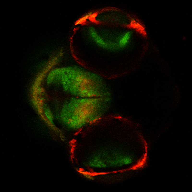
| Confocal image of brain of transgenic zebrafish (Dania rerio). Kaede protein – green is unconverted, red is photoconverted. Image courtesy of Robert Thorn, Creton Lab, Brown University. |