Questions? 800-523-5874 | [email protected]
- TEM Grids
- Prepmaster™ Specimen Preparation Robot
- TEM Window Grids
- Omniprobe Nanomanipulation Systems
- K-kit Wet "Liquid" TEM Kit
- Specimen Mounts
- SEM Specimen Holders
- Index and Finder SEM Grids
- SEM for Forensics
- SEM Sample Preparation Station Materials
- Cryogenic Personal Protection Equipment
- Cryo Dewars & Flasks
- Cryo-EM Grids & Grid Storage
- Cryogenic Vials & Racks
- Cryo-EM Vitrification Supplies
- Prepmaster™ Specimen Preparation Robot
- Laboratory Microwave Ovens
- LYNX II Automated Tissue Processor
- EMS Poly III
- Microtomes
- Tissue Slicers
- Heaters & Chillers
- SEM Cooling Stage
- Glow Discharge Systems
- Sputter Coaters & Carbon Coaters
- Stages
- Freeze Dryers
- Critical Point Dryers
- Cryo-SEM Preparation System
- Specimen Transfer Systems
- Decontaminators
- Desiccators
- Centrifuges
- Dry Baths
- Stirrers, Hot Plates
- Vortexers & Magnetic Mixers
- Rotators & Rockers
- Ovens & Incubators
- Vibration Isolation
- Air Sampling
- Vacuum Pumps
EMS Technical Data Sheets

Checking Fluorescence of Bacterial or Cell Cultures
EMS Catalog #SFA
We are occasionally asked about using the NIGHTSEA tools to check bacterial or cell cultures expressing GFP or other fluorophores. This has been done successfully. The informal report below was sent by a researcher trying out the BlueStar flashlight at a time before the Stereo Microscope Fluorescence Adapter was even a product. Now you can use NIGHTSEA gear to work at both the overview and detail levels. And if you have more than one color fluorophore you can also take advantage of the Dual Fluorescent Protein flashlight series.
The following text and images are exactly as supplied to us:
Project
Cell culture for in vitro transfection of NS20Y mouse cells with a construct that also expresses GFP.
Problem
I wanted to know 1) if my transfection was successful, and 2) I wanted to be able to track puromycin selection of GFP positive cells over a few weeks without having to take my cells to the core to see them under the fluorescent microscope every time.
Procedure
NIGHTSEA GFP Flashlight.
Conclusion
For a quick, qualitative assessment of success rate of transfection, the NIGHTSEA flashlight worked great. It also worked for tracking the increase in GFP positive cells over the time course of antibiotic selection (Fig 3). It is purely qualitative though. Using the flashlight, I actually thought I had a high number of puromycin resistant cells that were not GFP positive, but this was not the case. Once I looked at the cells under the microscope with a much higher intensity, I could confirm that most all my cells were indeed GFP positive (Fig 4). So I think the flashlight only picks up the highest fluorescing cells. For the cost and convenience, it is entirely worth it! The flashlight can also be used in the tissue culture hood to pick GFP positive cells to accelerate the cell selection process.
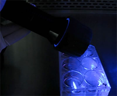 |
| Figure 1. BlueStar directed at wells |
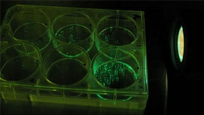 |
| Figure 2. BlueStar directed at wells, viewed through barrier filter glasses |
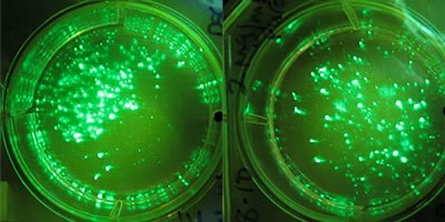 |
| Figure 3. Two wells illuminated by BlueStar and viewed through barrier filter glasses |
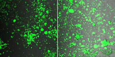 |
| Figure 4. Detail of wells under fluorescence microscope |