Questions? 800-523-5874 | [email protected]
- TEM Grids
- Prepmaster™ Specimen Preparation Robot
- TEM Window Grids
- Omniprobe Nanomanipulation Systems
- K-kit Wet "Liquid" TEM Kit
- Specimen Mounts
- SEM Specimen Holders
- Index and Finder SEM Grids
- SEM for Forensics
- SEM Sample Preparation Station Materials
- Cryogenic Personal Protection Equipment
- Cryo Dewars & Flasks
- Cryo-EM Grids & Grid Storage
- Cryogenic Vials & Racks
- Cryo-EM Vitrification Supplies
- Prepmaster™ Specimen Preparation Robot
- Laboratory Microwave Ovens
- LYNX II Automated Tissue Processor
- EMS Poly III
- Microtomes
- Tissue Slicers
- Heaters & Chillers
- SEM Cooling Stage
- Glow Discharge Systems
- Sputter Coaters & Carbon Coaters
- Stages
- Freeze Dryers
- Critical Point Dryers
- Cryo-SEM Preparation System
- Specimen Transfer Systems
- Decontaminators
- Desiccators
- Centrifuges
- Dry Baths
- Stirrers, Hot Plates
- Vortexers & Magnetic Mixers
- Rotators & Rockers
- Ovens & Incubators
- Vibration Isolation
- Air Sampling
- Vacuum Pumps
EMS Technical Data Sheets

Fluorescing Xenopus with Stereo Microscope Fluorescence Adapter
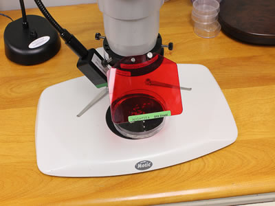
EMS Catalog #SFA
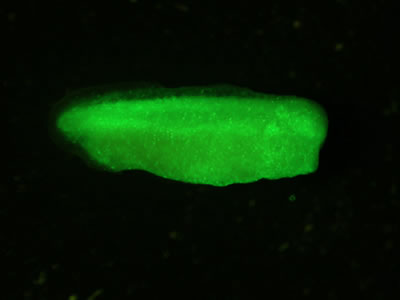
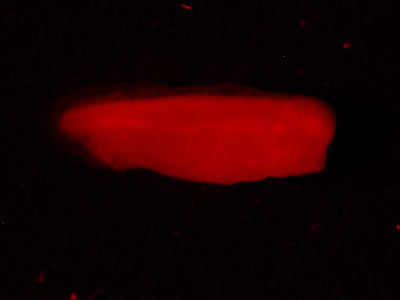
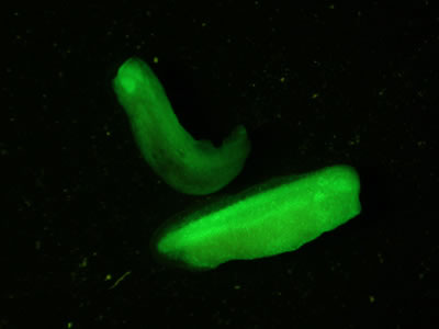
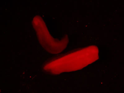
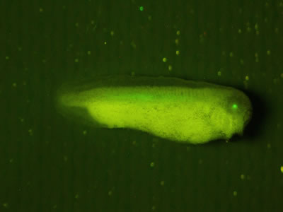
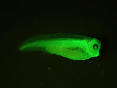
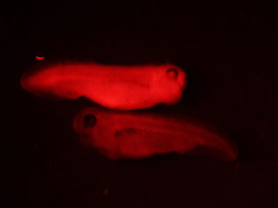
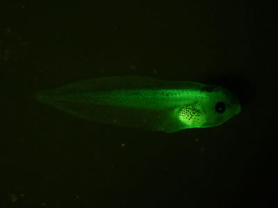
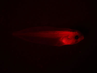
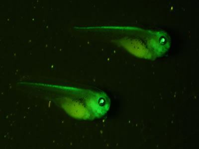
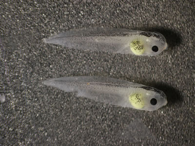
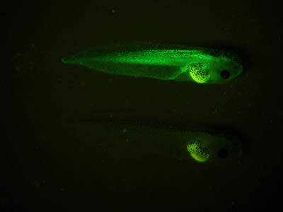
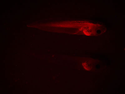
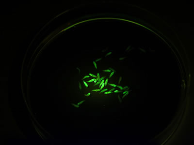
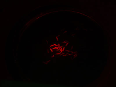
We had the opportunity to try out the NIGHTSEA Stereo Microscope Fluorescence Adapter at the National Xenopus Resource (NXR) at the Marine Biological Laboratory in Woods Hole, MA – special thanks to NXR Director and Bell Center Scientist Dr. Marko Horb and his postdoctoral scientist Dr. Matthew Salanga. The fluorescence adapter system worked great for visualizing all of the fluorescence, both injected and transgenic, in the specimens. In addition to seeing the fluorescence through the eyepieces, you could easily distinguish presence/absence and relative strength of expression just by looking through the filter shield. This mode can easily be used for selecting specimens.
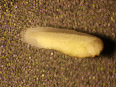
The varieties of Xenopus we looked at were:
- Stage 29-30 X. laevis with messenger RNA injected ubiquitous GFP and membrane RFP
- Stage 37-38 X. laevis with messenger RNA injected ubiquitous GFP and membrane RFP
- Stage 46 X. laevis with messenger RNA injected ubiquitous GFP and membrane RFP
- Stage 41 X. tropicalis transgenic OTX-GFP eyes
- Stage 32 X. laevis transgenic GFP col2a1 early expression in eyes and back
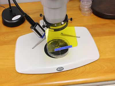
All of the specimen photographs below were taken with a Canon Rebel T2i camera mounted on a Motic trinocular stereo microscope with the NIGHTSEA Stereo Microscope Fluorescence Adapter for illumination and filtering.