Questions? 800-523-5874 | [email protected]
- TEM Grids
- Prepmaster™ Specimen Preparation Robot
- TEM Window Grids
- Omniprobe Nanomanipulation Systems
- K-kit Wet "Liquid" TEM Kit
- Specimen Mounts
- SEM Specimen Holders
- Index and Finder SEM Grids
- SEM for Forensics
- SEM Sample Preparation Station Materials
- Cryogenic Personal Protection Equipment
- Cryo Dewars & Flasks
- Cryo-EM Grids & Grid Storage
- Cryogenic Vials & Racks
- Cryo-EM Vitrification Supplies
- Prepmaster™ Specimen Preparation Robot
- Laboratory Microwave Ovens
- LYNX II Automated Tissue Processor
- EMS Poly III
- Microtomes
- Tissue Slicers
- Heaters & Chillers
- SEM Cooling Stage
- Glow Discharge Systems
- Sputter Coaters & Carbon Coaters
- Stages
- Freeze Dryers
- Critical Point Dryers
- Cryo-SEM Preparation System
- Specimen Transfer Systems
- Decontaminators
- Desiccators
- Centrifuges
- Dry Baths
- Stirrers, Hot Plates
- Vortexers & Magnetic Mixers
- Rotators & Rockers
- Ovens & Incubators
- Vibration Isolation
- Air Sampling
- Vacuum Pumps
AURION ImmunoGold Reagents - Micrographs

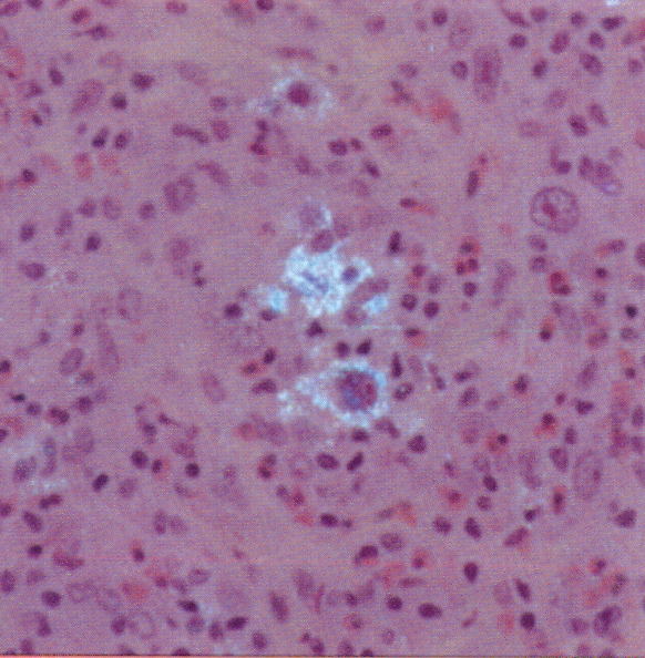
Routine paraffin section of Hodgkin lymphoma stained for CD 15. Reed-Sternberg cells show positive staining in the cytoplasm. Products used:
- Mouse monoclonal CD 15
- GAM IgG GP-US
- R-Gent
 Combination regular light microscopy and epi-polarization microscopy |
 Combination regular light microscopy and epi-polarization microscopy |
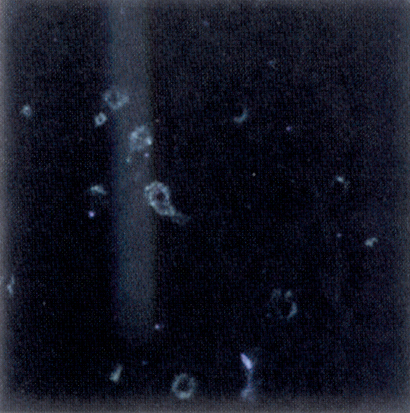 Epi-polarization microscopy |
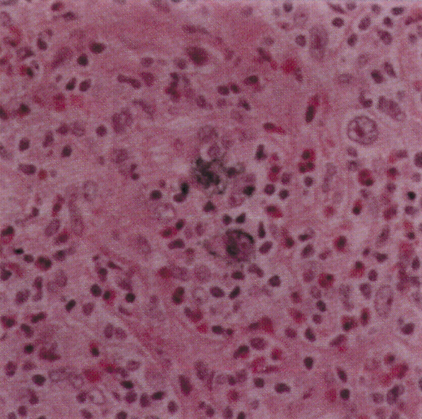
Products used: Rabbit polyclonal to alpha-amylase, GAR-GP-Ultrasmall R-Gent
 Combination regular light microscopy and epi-polarization microscopy |
 Combination regular light microscopy and epi-polarization microscopy |
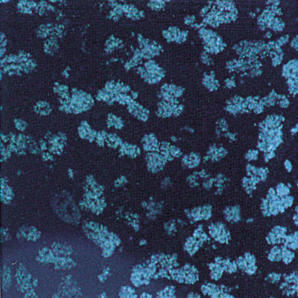 Epi-polarization microscopy |
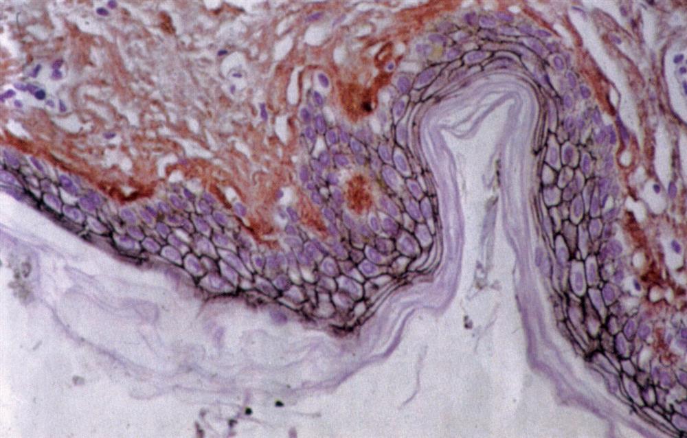 Immunogold Silver Staining of E-cadherin on a paraffin section of human skin.
Courtesy of R. Moella, Dept. of Exp. Path., EUR, The Netherlands.
• Mouse monoclonal anti E-cadherin
• GAM lgG UltraSmall
• Aurion R-Gent SE-LM
 |
 |
||
|
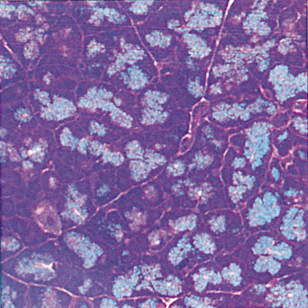
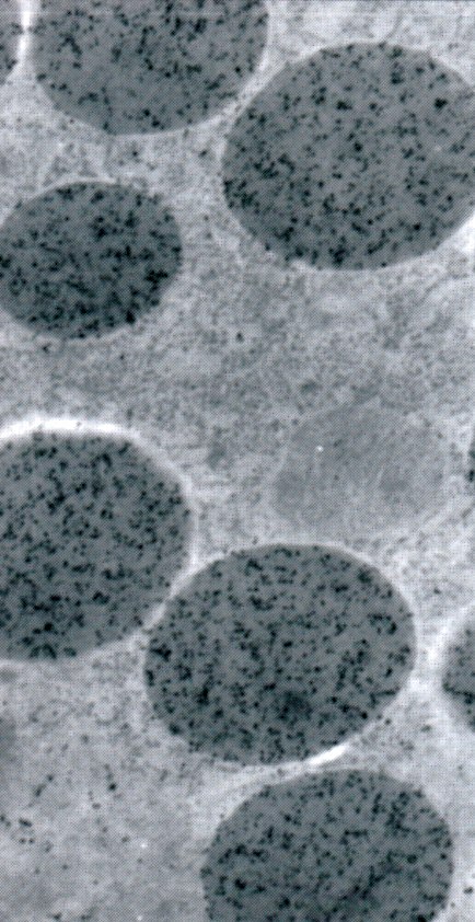
Immunogold silver staining of alpha-amylase on Lowicryl HM20 section of rat pancreas.
|
||
| Pre-embedding Immunogold Labeling of Huntingtin Interacting Protein 3 in Mouse Brain using Aurion GAR Fab-US and Aurion SEEM. Courtesy of Ms Hong Yi, Emory University, Atlanta GA |
 |
||
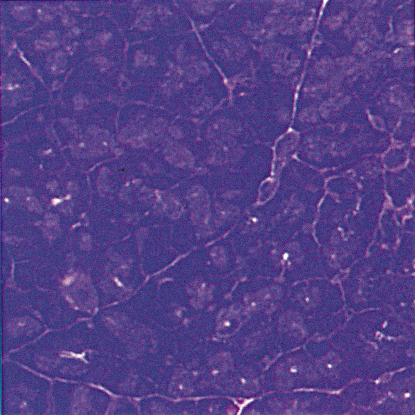
| IGSS of tubulin on coverslip culture of PtK2 cells Courtesy of Peter van de Plas, Aurion Costerweg 5, The Netherlands. |  |
||
AURION R-Gent SE-EM Application Example
 |
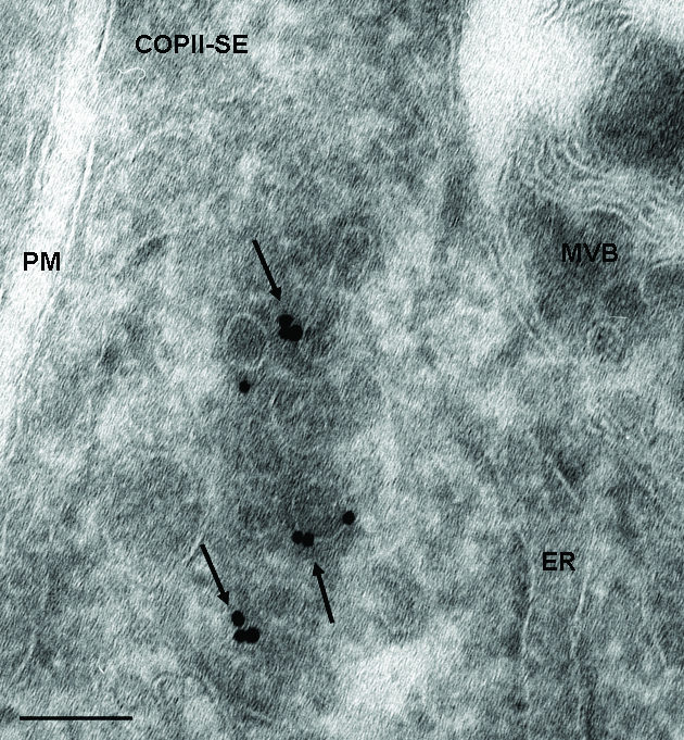 |
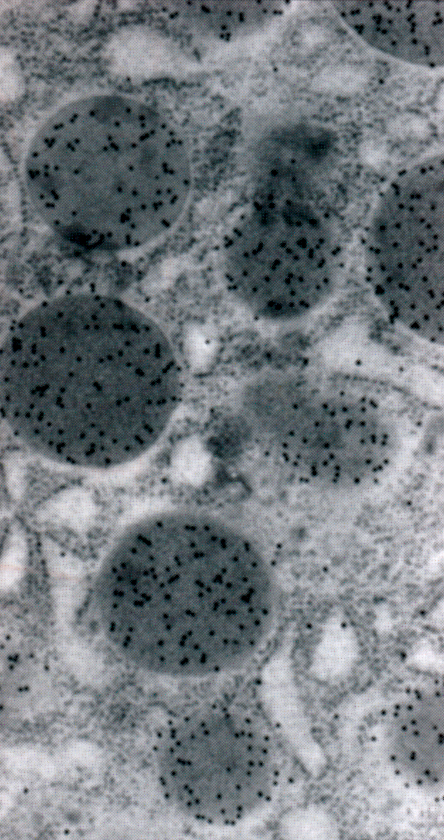
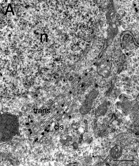
ER exit site in 60 nm-thin cryosection of Hepg2 cells, labeled for COPII (primary antibody against sec23 was obtained by ABR) and detected with Fab-goat-anti-rabbit, conjugated to ultra-small gold, silver enhanced for 30 minutes (from Aurion). The arrows point to labeled COPII-coats on vesicular and tubular membranes, which are located close to the ER. The information of a thin section is not sufficient to conclude how the membranes are related to each other- if they are still connected to the ER, or if they are free. Therefore we performed 3D electron tomography on 400nm thick cryosections, which were labeled similar for COPII (see next picture). |
||||
|
|
||||||
 |
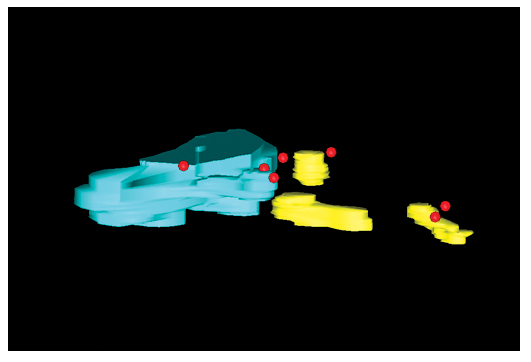 |
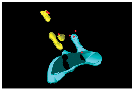
2 views of a model of a COPII-labeled ER-exit site, resolved from 400nm thick cryo-sections of Hepg2 cells, labeled like described for the ultrathin section before. Note that the labeling for COPII is assessable throughout the section. Courtesy of: Dagmar Zeuschner, Judith Klumperman (Department of Cell Biology, UMC Utrecht, The Netherlands) and Willie Geerts, Abraham Koster (Molecular Cell Biology, Utrecht University, The Netherlands) |
||||
| ER = light blue, Free membrane carriers of vesicular and tubular shape, partially labeled for COPII = yellow, COPII = silver enhanced-red |
||||||